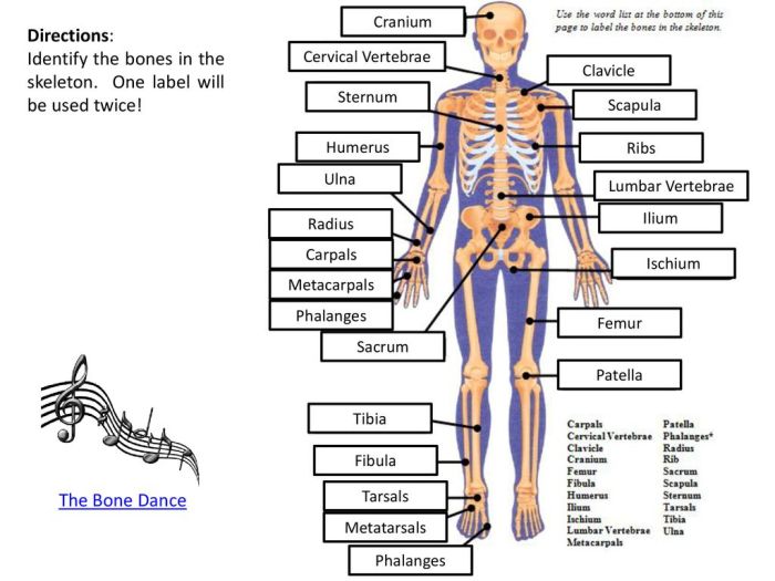
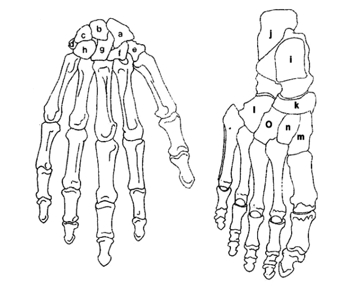
Label the carpals and tarsals – Labeling the carpals and tarsals takes center stage as we delve into the intricate realm of the human skeletal system. These bones, found in the wrist and foot respectively, play pivotal roles in our everyday movements and deserve our utmost attention.
Join us on an enlightening journey as we unravel the mysteries surrounding these fascinating structures, exploring their names, locations, and functions with unparalleled clarity.
Label the Carpals

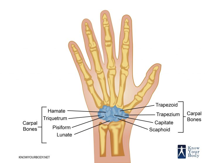
The carpal bones are a group of eight small bones that form the wrist. They are arranged in two rows, with four bones in each row.
The carpals and tarsals are small bones that form the wrist and ankle, respectively. They are important for movement and stability. While we’re on the topic of interesting facts, have you ever wondered if Jehovah’s Witnesses eat pork? Do Jehovah’s Witnesses eat pork ? It’s a common question, and the answer might surprise you.
Back to the carpals and tarsals, it’s fascinating how these tiny bones play such a vital role in our bodies.
The proximal row of carpal bones, from lateral to medial, includes the scaphoid, lunate, triquetrum, and pisiform bones. The distal row of carpal bones, from lateral to medial, includes the trapezium, trapezoid, capitate, and hamate bones.
Name, Location, and Function of Carpal Bones
| Name | Location | Function |
|---|---|---|
| Scaphoid | Proximal row, lateral | Articulates with the radius and ulna proximally, and the trapezium, trapezoid, and capitate distally |
| Lunate | Proximal row, middle | Articulates with the radius and ulna proximally, and the capitate and hamate distally |
| Triquetrum | Proximal row, medial | Articulates with the ulna proximally, and the pisiform, hamate, and lunate distally |
| Pisiform | Proximal row, anterior | Articulates with the triquetrum proximally, and the hamate and capitate distally |
| Trapezium | Distal row, lateral | Articulates with the scaphoid and trapezoid proximally, and the first metacarpal distally |
| Trapezoid | Distal row, middle | Articulates with the scaphoid and trapezium proximally, and the second metacarpal distally |
| Capitate | Distal row, middle | Articulates with the scaphoid, lunate, and hamate proximally, and the second, third, and fourth metacarpals distally |
| Hamate | Distal row, medial | Articulates with the lunate, triquetrum, and pisiform proximally, and the fourth and fifth metacarpals distally |
Label the Tarsals

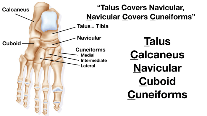
The tarsals are a group of seven bones that form the ankle joint. They are located between the tibia and fibula (the two bones of the lower leg) and the metatarsals (the five bones of the foot). The tarsals help to support the weight of the body and allow for movement of the foot.
Tarsal Bones
The seven tarsal bones are:
- Talus:The talus is the largest tarsal bone. It is located at the top of the ankle joint and articulates with the tibia and fibula. The talus helps to transmit weight from the leg to the foot.
- Calcaneus:The calcaneus is the heel bone.
It is the largest and strongest tarsal bone. The calcaneus helps to support the weight of the body and provides leverage for the muscles of the foot.
- Navicular:The navicular is a small bone located on the medial side of the foot.
It articulates with the talus, calcaneus, and three cuneiform bones. The navicular helps to support the arch of the foot.
- Medial cuneiform:The medial cuneiform is the smallest of the three cuneiform bones. It is located on the medial side of the foot and articulates with the navicular and first metatarsal.
The medial cuneiform helps to support the arch of the foot.
- Intermediate cuneiform:The intermediate cuneiform is located between the medial and lateral cuneiform bones. It articulates with the navicular and second metatarsal. The intermediate cuneiform helps to support the arch of the foot.
- Lateral cuneiform:The lateral cuneiform is the largest of the three cuneiform bones. It is located on the lateral side of the foot and articulates with the navicular and third metatarsal. The lateral cuneiform helps to support the arch of the foot.
- Cuboid:The cuboid is a small bone located on the lateral side of the foot. It articulates with the calcaneus, lateral cuneiform, and fourth and fifth metatarsals. The cuboid helps to support the arch of the foot.
Compare the Carpals and Tarsals

The carpals and tarsals are two sets of small bones located in the wrist and ankle, respectively. They play a crucial role in providing stability, mobility, and shock absorption in these joints.
Number of Bones
The carpus consists of eight bones, while the tarsus comprises seven bones.
Location, Label the carpals and tarsals
The carpals are located between the radius and ulna (forearm bones) and the metacarpals (hand bones). The tarsals are positioned between the tibia and fibula (leg bones) and the metatarsals (foot bones).
Function
Both the carpals and tarsals provide structural support and allow for movement in their respective joints. The carpals facilitate hand movements such as flexion, extension, and rotation. The tarsals, on the other hand, enable ankle movements like plantarflexion, dorsiflexion, and inversion/eversion.
Mobility
The carpals exhibit greater mobility compared to the tarsals. This increased mobility allows for a wider range of hand movements, including fine motor skills.
| Feature | Carpals | Tarsals |
|---|---|---|
| Number of Bones | 8 | 7 |
| Location | Wrist | Ankle |
| Function | Structural support, hand movement | Structural support, ankle movement |
| Mobility | Greater | Less |
Illustrate the Carpal and Tarsal Bones: Label The Carpals And Tarsals

The carpal and tarsal bones are two sets of bones that make up the wrist and ankle joints, respectively. They are both made up of eight small bones that are arranged in two rows. The carpal bones are located between the forearm and the metacarpal bones of the hand, while the tarsal bones are located between the leg and the metatarsal bones of the foot.
A Diagram of the Carpal Bones in the Wrist
The carpal bones are arranged in two rows, with four bones in each row. The proximal row of carpal bones consists of the scaphoid, lunate, triquetrum, and pisiform bones. The distal row of carpal bones consists of the trapezium, trapezoid, capitate, and hamate bones.
- The scaphoid bone is located on the thumb side of the wrist and is shaped like a boat.
- The lunate bone is located next to the scaphoid bone and is shaped like a crescent moon.
- The triquetrum bone is located on the little finger side of the wrist and is shaped like a triangle.
- The pisiform bone is a small, pea-shaped bone that is located on the palmar side of the wrist.
- The trapezium bone is located on the thumb side of the wrist and is shaped like a trapezoid.
- The trapezoid bone is located next to the trapezium bone and is shaped like a trapezoid.
- The capitate bone is located in the center of the wrist and is shaped like a head.
- The hamate bone is located on the little finger side of the wrist and is shaped like a hook.
An Image of the Tarsal Bones in the Foot
The tarsal bones are arranged in two rows, with three bones in the proximal row and five bones in the distal row. The proximal row of tarsal bones consists of the talus, calcaneus, and navicular bones. The distal row of tarsal bones consists of the cuboid, lateral cuneiform, intermediate cuneiform, and medial cuneiform bones.
- The talus bone is located at the top of the ankle joint and is shaped like a dome.
- The calcaneus bone is the largest tarsal bone and is located at the back of the heel.
- The navicular bone is located between the talus and the three cuneiform bones.
- The cuboid bone is located on the lateral side of the foot and is shaped like a cube.
- The lateral cuneiform bone is located on the lateral side of the foot and is shaped like a wedge.
- The intermediate cuneiform bone is located in the middle of the foot and is shaped like a wedge.
- The medial cuneiform bone is located on the medial side of the foot and is shaped like a wedge.
FAQ Insights
What is the difference between carpals and tarsals?
Carpals are the bones of the wrist, while tarsals are the bones of the foot.
How many carpals are there?
There are 8 carpals.
How many tarsals are there?
There are 7 tarsals.